
Page 156 - Synthesis of Functional Nanoparticles Using an Atmospheric Pressure Microplasma Process - LiangLiang Lin
P. 156
Chapter 8
To get an overview of the obtain products and to correlate processing parameters with their morphology, TEM characterization was performed By comparing TEM images of the products (Figure 8.4), it is noted that Ag nanoparticles produced by Ag(NH3)2+ reduction without dispersant have a narrower particle size distribution than those derived from Ag+ reduction in the same condition. Similar to the Ag+ reaction system, dispersant can greatly improve the particle uniformity and control the particle size in the Ag(NH3)2+ reaction system. The average size of AgNPs from Ag(NH3)2+ and Ag+ with dispersant was 15.2 and 35.9 nm, respectively.
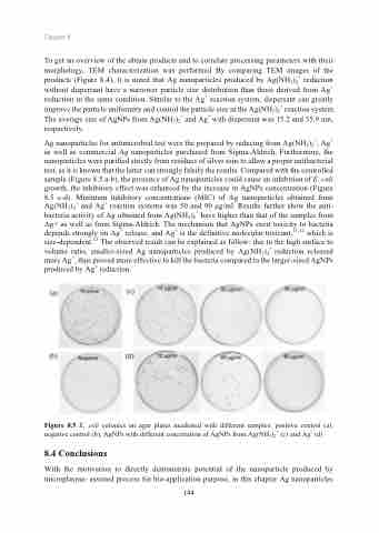
Ag nanoparticles for antimicrobial test were the prepared by reducing from Ag(NH3)2+, Ag+ as well as commercial Ag nanoparticles purchased from Sigma-Aldrich. Furthermore, the nanoparticles were purified strictly from residues of silver ions to allow a proper antibacterial test, as it is known that the latter can strongly falsify the results. Compared with the controlled sample (Figure 8.5 a-b), the presence of Ag nanoparticles could cause an inhibition of E. coli growth, the inhibitory effect was enhanced by the increase in AgNPs concentration (Figure 8.5 c-d). Minimum inhibitory concentrations (MIC) of Ag nanoparticles obtained from Ag(NH3)2+ and Ag+ reaction systems was 50 and 90 μg/ml. Results further show the anti- bacteria activity of Ag obtained from Ag(NH3)2+ have higher than that of the samples from Ag+ as well as from Sigma-Aldrich. The mechanism that AgNPs exert toxicity to bacteria depends strongly on Ag+ release, and Ag+ is the definitive molecular toxicant,11,12 which is size-dependent.13 The observed result can be explained as follow: due to the high surface to volume ratio, smaller-sized Ag nanoparticles produced by Ag(NH3)2+ reduction released more Ag+, thus proved more effective to kill the bacteria compared to the larger-sized AgNPs produced by Ag+ reduction.
Figure 8.5 E. coli colonies on agar plates incubated with different samples: positive control (a), negative control (b), AgNPs with different concertation of AgNPs from Ag(NH3)2+ (c) and Ag+ (d)
8.4 Conclusions
With the motivation to directly demonstrate potential of the nanoparticle produced by microplasma- assisted process for bio-application purpose, in this chapter Ag nanoparticles
144
