
Page 43 - Physico-Chemical Niche Conditions for Bone Cells
P. 43
myoblasts, the glycocalyx is critically required for PFF-induced NO production [15]. However, whether the glycocalyx plays a role in mechanosensing by MuSCs is unknown. Local stretching of the cell membrane will likely activate the stretch-activated calcium channels, which could elicit several signaling pathways. Further research is required to disentangle the functional role of the glycocalyx and the deformation of the cell membrane, cytoskeleton, and nucleus on signaling pathways and changes in gene expression.
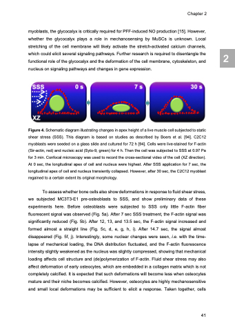
Figure 4. Schematic diagram illustrating changes in apex height of a live muscle cell subjected to static shear stress (SSS). This diagram is based on studies as described by Boers et al. [94]. C2C12 myoblasts were seeded on a glass slide and cultured for 72 h [94]. Cells were live-stained for F-actin (Sir-actin, red) and nucleic acid (Syto-9, green) for 4 h. Then the cell was subjected to SSS at 0.97 Pa for 3 min. Confocal microscopy was used to record the cross-sectional video of the cell (XZ direction). At 0 sec, the longitudinal apex of cell and nucleus were highest. After SSS application for 7 sec, the longitudinal apex of cell and nucleus transiently collapsed. However, after 30 sec, the C2C12 myoblast regained to a certain extent its original morphology.
To assess whether bone cells also show deformations in response to fluid shear stress, we subjected MC3T3-E1 pre-osteoblasts to SSS, and show preliminary data of these experiments here. Before osteoblasts were subjected to SSS only little F-actin fiber fluorescent signal was observed (Fig. 5a). After 7 sec SSS treatment, the F-actin signal was significantly reduced (Fig. 5b). After 12, 13, and 13.5 sec, the F-actin signal increased and formed almost a straight line (Fig. 5c, d, e, g, h, i). After 14.7 sec, the signal almost disappeared (Fig. 5f, j). Interestingly, some nuclear changes were seen, i.e. with the time- lapse of mechanical loading, the DNA distribution fluctuated, and the F-actin fluorescence intensity slightly weakened as the nucleus was slightly compressed, showing that mechanical loading affects cell structure and (de)polymerization of F-actin. Fluid shear stress may also affect deformation of early osteocytes, which are embedded in a collagen matrix which is not completely calcified. It is expected that such deformations will become less when osteocytes mature and their niche becomes calcified. However, osteocytes are highly mechanosensitive and small local deformations may be sufficient to elicit a response. Taken together, cells
Chapter 2
41
2
