
Page 42 - Physico-Chemical Niche Conditions for Bone Cells
P. 42
Physicochemical niche conditions and mechanosensing
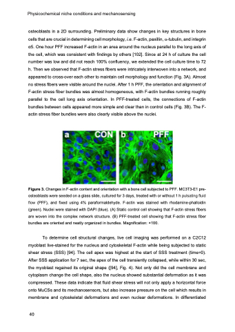
osteoblasts in a 2D surrounding. Preliminary data show changes in key structures in bone cells that are crucial in determining cell morphology, i.e. F-actin, paxillin, a-tubulin, and integrin α5. One hour PFF increased F-actin in an area around the nucleus parallel to the long axis of the cell, which was consistent with findings by others [102]. Since at 24 h of culture the cell number was low and did not reach 100% confluency, we extended the cell culture time to 72 h. Then we observed that F-actin stress fibers were intricately interwoven into a network, and appeared to cross-over each other to maintain cell morphology and function (Fig. 3A). Almost no stress fibers were visible around the nuclei. After 1 h PFF, the orientation and alignment of F-actin stress fiber bundles was almost homogeneous, with F-actin bundles running roughly parallel to the cell long axis orientation. In PFF-treated cells, the connections of F-actin bundles between cells appeared more simple and clear than in control cells (Fig. 3B). The F- actin stress fiber bundles were also clearly visible above the nuclei.
Figure 3. Changes in F-actin content and orientation with a bone cell subjected to PFF. MC3T3-E1 pre- osteoblasts were seeded on a glass slide, cultured for 3 days, treated with or without 1 h pulsating fluid flow (PFF), and fixed using 4% paraformaldehyde. F-actin was stained with rhodamine-phalloidin (green). Nuclei were stained with DAPI (blue). (A) Static control cell showing that F-actin stress fibers are woven into the complex network structure. (B) PFF-treated cell showing that F-actin stress fiber bundles are oriented and neatly organized in bundles. Magnification: ×100.
To determine cell structural changes, live cell imaging was performed on a C2C12 myoblast live-stained for the nucleus and cytoskeletal F-actin while being subjected to static shear stress (SSS) [94]. The cell apex was highest at the start of SSS treatment (time=0). After SSS application for 7 sec, the apex of the cell transiently collapsed, while within 30 sec, the myoblast regained its original shape ([94], Fig. 4). Not only did the cell membrane and cytoplasm change the cell shape, also the nucleus showed substantial deformation as it was compressed. These data indicate that fluid shear stress will not only apply a horizontal force onto MuCSs and its mechanosensors, but also increase pressure on the cell which results in membrane and cytoskeletal deformations and even nuclear deformations. In differentiated
40
