
Page 151 - 89Zr-Immuno-PET:Towards a Clinical Tool to Guide Antibody-based Therapy in Cancer
P. 151
89Zr-rituximab-PET in lymphoma
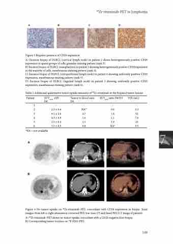
Figure 3 Biopsies: presence of CD20 expression
A) Excision biopsy of DLBCL (cervical lymph node) in patient 2 shows heterogeneously positive CD20 expression in sparse groups of cells, granular staining pattern (rank 3).
B) Excision biopsy of DLBCL (nasopharynx) in patient 5 showing heterogeneously positive CD20 expression in the majority of cells, membranous staining pattern (rank 4).
C) Excision biopsy of DLBCL (retroperitoneal lymph node) in patient 4 showing uniformly positive CD20 expression, membranous staining pattern (rank 5).
D) Excision biopsy of DLBCL (inguinal lymph node) in patient 3 showing uniformly positive CD20 expression, membranous staining pattern (rank 6).
Table 2 Additional quantitative tumor uptake measures of 89Zr-rituximab in the biopsied tumor lesions
Patient SUVmean ±SD Tumor to blood ratio SUVpeak ratio D6/D3 VOI (mL) D6 D6
1----
2 3 4 5 6
*NA = not available
2.3 ± 0.4 9.1 ± 2.6 4.3 ± 0.9 2.5 ± 0.4 3.5 ± 0.5
NA* 0.6 4.7 1.4 1.6 1.1 1.1 1.0 0.8 NA*
5.3 32 7.6 25 3.5
7
Figure 4 No tumor uptake on 89Zr-rituximab-PET, concordant with CD20 expression in biopsy. Axial images: from left to right attenuation corrected PET, low dose CT and fused PET/CT image of patient1.
A) 89Zr-rituximab-PET shows no tumor uptake concordant with a CD20-negative liver biopsy. B) Corresponding tumor location on 18F-FDG-PET.
149
