
Page 150 - 89Zr-Immuno-PET:Towards a Clinical Tool to Guide Antibody-based Therapy in Cancer
P. 150
Chapter 7
All sites of positive tumor uptake identified at D6 were also observed at D3, whereas no tumor uptake was identified on D0. At D6 on average 27.6% ± 5.7% ID of 89Zr-rituximab was still circulating in blood pool. The total activity at D6 as derived from plasma samples was not significantly different from the whole blood samples (n=5). Image derived and venous sampled whole blood activity concentrations were correlated with an R2 of 0.98 and slope of 0.85.
All biopsied tumor sites showed uptake of 18F-FDG and DLBCL localization was confirmed by pathology. IHC was negative for CD20 expression in patient 1 and 6 (Figure 2), and positive in the other patients (Figure 3). Patients were ranked based on the intensity level and pattern of CD20 expression.
Tumor uptake of 89Zr-rituximab and CD20 expression on IHC were concordant in five patients: for patient 1, both were negative (Figure 4 and 2A), for the other four patients visible tumor uptake was concordant with CD20-positive biopsies. Intense visual tumor uptake of 89Zr-rituximab on PET was observed in patient 3, corresponding with uniformly positive CD20 expression on IHC (Figure 5 and 3D). SUVpeak for this tumor lesion on D6 was 12.8. CD20 expression on IHC was also observed in patient 2 (Figure 3A), 4 (Figure 3B) and 5 (Figure 3c), concordant with tumor uptake of 89Zr-rituximab. SUVpeak for these tumor lesions on D6 was 3.2, 5.4, 3.4, respectively. In patient 6 tumor uptake of 89Zr-rituximab was observed (SUVpeak on D6 = 3.8) (Figure 6) while a core needle biopsy was CD20 negative (Figure 2B). Tumor uptake over time (SUVpeak D6/D3 ratio) was calculated and ranged between 0.6 and 1.4. See Table 2 for additional PET uptake measures.
Overall, a positive correlation was observed between tumor uptake of 89Zr- rituximab, measured as SUVpeak, and the CD20 expression ranking (rs = 0.83, p=0.04, n = 6).
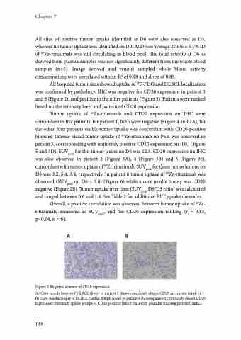
Figure 2 Biopsies: absence of CD20 expression
A) Core-needle biopsy of DLBCL (liver) in patient 1 shows completely absent CD20 expression (rank 1). B) Core-needle biopsy of DLBCL (axillar lymph node) in patient 6 showing almost completely absent CD20 expression: extremely sparse groups of CD20-positive tumor cells with granular staining pattern (rank2).
148
