
Page 132 - 89Zr-Immuno-PET:Towards a Clinical Tool to Guide Antibody-based Therapy in Cancer
P. 132
Chapter 6
n=103). The range in interobserver variability between VOI within a single patient was 0 to 2.3 (median 0.6, n=22) for patient 2 (89Zr-rituximab at D6). Interobserver variability (SEM) at D6 for the remaining five 89Zr-rituximab patients ranged from 0.1 to 1.4 (median 0.3, n=8). Thus, as interobserver variability was higher within a single patient than between patients, a VOI-based analysis was performed.
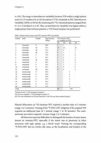
Table 1 Patient characteristics and 89Zr-immuno-PET scan details
Patient mAb
1 rituximab
2 rituximab
3 rituximab
4 rituximab
5 rituximab
6 cetuximab
7 cetuximab
8 cetuximab
9 cetuximab
10 cetuximab
11 cetuximab
12 trastuzumab
13 trastuzumab
14 trastuzumab
15 trastuzumab
16 trastuzumab
17 trastuzumab
18 trastuzumab
Total
Gender Injected dose (MBq)
F 69.8 M 75.3 M 79.2 M 75.0 F 75.6 F 36.7 M 35.6 F 36.2 F 36.5 F 35.5 M 38.1 F 35.0 F 38.2 F 35.8 F 37.3 F 38.3 F 35.3 F 37.0
89Zr-immuno-PET (n= number of VOI)
D0 D3 D4 D6
1
22
2
0*
6
2
2
2
1
2
0†
-
-
-
-
-
-
-
40
1 - 22 - 2 - 1 - 0 - 2 - 2 - 2 - 1 - 0 - 0 - - 5 - 4 - 5 - 4 - 5 - 2 - 3
1 22 2 1 6 2 2 2 1 2 1 -
- - - - - -
103
*Technical error: 1 VOI missing for patient 4.
†No D0 scan available: 1 VOI missing for patient 11.
D0 VOI were delineated on D6 and imported to the D0 scan (data marked in grey).
Manual delineation on 89Zr-immuno-PET required a median time of 2 minutes (range 1 to 5 minutes). Viewing of the 18F-FDG-PET /adaption of the original VOI required an additional time of 1 minute (range 1 to 30 minutes). The semi- automatic procedure required 1 minute (range 1 to 5 minutes).
All observers reported difficulties to distinguish the borders of some tumor lesions on immuno-PET, especially if the tumor was in proximity to other structures with high uptake, e.g. a blood vessel. Viewing the corresponding 18F-FDG-PET did not resolve this issue, as the localization and borders of the
130
