
Page 39 - Advanced concepts in orbital wall fractures
P. 39
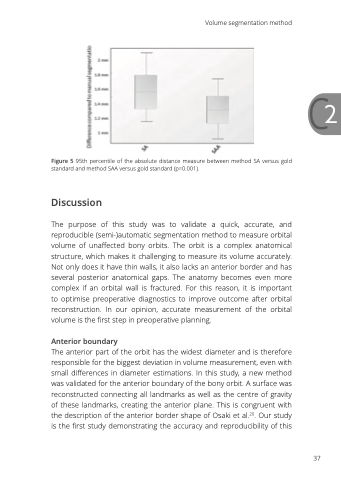
                                C2 Figure 5 95th percentile of the absolute distance measure between method SA versus gold
standard and method SAA versus gold standard (p=0.001).
Discussion
The purpose of this study was to validate a quick, accurate, and reproducible (semi-)automatic segmentation method to measure orbital volume of unaffected bony orbits. The orbit is a complex anatomical structure, which makes it challenging to measure its volume accurately. Not only does it have thin walls, it also lacks an anterior border and has several posterior anatomical gaps. The anatomy becomes even more complex if an orbital wall is fractured. For this reason, it is important to optimise preoperative diagnostics to improve outcome after orbital reconstruction. In our opinion, accurate measurement of the orbital volume is the first step in preoperative planning.
Anterior boundary
The anterior part of the orbit has the widest diameter and is therefore responsible for the biggest deviation in volume measurement, even with small differences in diameter estimations. In this study, a new method was validated for the anterior boundary of the bony orbit. A surface was reconstructed connecting all landmarks as well as the centre of gravity of these landmarks, creating the anterior plane. This is congruent with the description of the anterior border shape of Osaki et al.20. Our study is the first study demonstrating the accuracy and reproducibility of this
Volume segmentation method
  37
