
Page 34 - Advanced concepts in orbital wall fractures
P. 34
Chapter 2
Gold standard
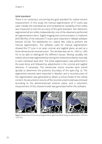
There is no consensus concerning the gold standard for orbital volume measurement. In this study, the manual segmentation of CT scans was used. Initially, the interobserver and intraobserver variability of ten orbits was measured to test the accuracy of this gold standard. Two observers segmented all ten orbits independently; one of the observers performed all segmentations twice. Digital imaging and communication in medicine (DICOM) files of the selected CT scans were imported in Matlab software (version 2012b; The Mathworks Inc., Natick, MA, USA) to perform the manual segmentation. The software used for manual segmentation showed the CT scan in an axial, coronal, and sagittal plane, as well as a three-dimensional reconstruction. The window was set to −200 to +200 HU to be able to distinguish the different tissues. Moving caudally, the orbital volume was segmented by tracing the orbital boundaries manually in each individual axial slice. The initial segmentation was performed in the axial slices and followed by adjustments in the coronal and sagittal direction, if necessary. The extraocular rectus muscles were traced apically to determine the posterior boundary of the apex (Fig. 2). The segmented volumes were imported in Maxilim, and a reconstruction of the segmentation was generated to obtain a virtual model of the orbital content. Excess anterior volume of the reconstructed model was removed according to the aforementioned anterior plane, and a volumetric measurement of this cleaned model was generated within the software.
Figure 2 Axial slices of the manual segmentation (gold standard) using a small window (−200 and +200 HU).
32
