
Page 33 - Advanced concepts in orbital wall fractures
P. 33
Anatomical boundaries
To calculate a volume in general, a virtually enclosed space is needed.
In order to be able to compare the different methods, the orbital
boundaries need to be defined first. The anterior boundary is reported C to be difficult to define20. In this study, interobserver agreement for the 2 anterior boundary was measured using the following method.
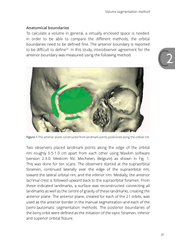
Figure 1 The anterior plane constructed from landmark points positioned along the orbital rim.
Two observers placed landmark points along the edge of the orbital rim roughly 0.5-1.0 cm apart from each other using Maxilim software (version 2.3.0; Medicim NV, Mechelen, Belgium) as shown in Fig. 1. This was done for ten scans. The observers started at the supraorbital foramen, continued laterally over the edge of the supraorbital rim, toward the lateral orbital rim, and the inferior rim. Medially, the anterior lacrimal crest is followed upward back to the supraorbital foramen. From these indicated landmarks, a surface was reconstructed connecting all landmarks as well as the centre of gravity of these landmarks, creating the anterior plane. The anterior plane, created for each of the 21 orbits, was used as the anterior border in the manual segmentation and each of the (semi-)automatic segmentation methods. The posterior boundaries of the bony orbit were defined as the initiation of the optic foramen, inferior and superior orbital fissure.
Volume segmentation method
31
