
Page 59 - Effects of radiotherapy and hyperbaric oxygen therapy on oral microcirculation Renee Helmers
P. 59
HBOT and vascular regeneration in oral mucosal flaps
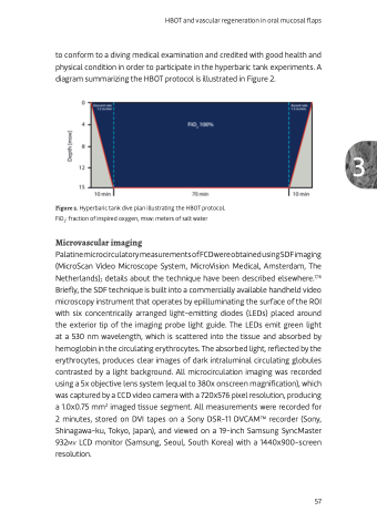
to conform to a diving medical examination and credited with good health and physical condition in order to participate in the hyperbaric tank experiments. A diagram summarizing the HBOT protocol is illustrated in Figure 2.
Figure 2. Hyperbaric tank dive plan illustrating the HBOT protocol. FiO2: fraction of inspired oxygen, msw: meters of salt water
Microvascular imaging
PalatinemicrocirculatorymeasurementsofFCDwereobtainedusingSDFimaging (MicroScan Video Microscope System, MicroVision Medical, Amsterdam, The Netherlands); details about the technique have been described elsewhere.7,16 Briefly, the SDF technique is built into a commercially available handheld video microscopy instrument that operates by epiilluminating the surface of the ROI with six concentrically arranged light-emitting diodes (LEDs) placed around the exterior tip of the imaging probe light guide. The LEDs emit green light at a 530 nm wavelength, which is scattered into the tissue and absorbed by hemoglobin in the circulating erythrocytes. The absorbed light, reflected by the erythrocytes, produces clear images of dark intraluminal circulating globules contrasted by a light background. All microcirculation imaging was recorded using a 5x objective lens system (equal to 380x onscreen magnification), which was captured by a CCD video camera with a 720x576 pixel resolution, producing a 1.0x0.75 mm2 imaged tissue segment. All measurements were recorded for 2 minutes, stored on DVI tapes on a Sony DSR-11 DVCAMTM recorder (Sony, Shinagawa-ku, Tokyo, Japan), and viewed on a 19-inch Samsung SyncMaster 932mv LCD monitor (Samsung, Seoul, South Korea) with a 1440x900-screen resolution.
57
3
