
Page 32 - Effects of radiotherapy and hyperbaric oxygen therapy on oral microcirculation Renee Helmers
P. 32
Chapter 2
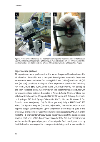
Figure 1. MicroScan mounted on top of a modified micromanipulator (A) and the setup of the MicroScan apparatus intraorally (B) targeting the right sublingual mucosa (probe the left side of the image) and the endotracheal tube oriented towards the left side of the oral cavity (on the right side of the image).
Experimental protocol
All experiments were performed at the same designated location inside the HB chamber. Since this was a two-part investigation, sequential hyperoxic experiments were conducted first during NB (1 atm [1.0 bar]) and then HB (2.5 atm [2.5 bar]) conditions. Each part of the experiment consisted of switching FiO2 from 21% to 55%, 100%, and back to 21% once every 10 min during NB and then repeated at HB. An overview of the experimental procedures with corresponding time points is illustrated in Figure 2. Serial 0.5 mL of blood was withdrawn into heparinized (Heparin LEO®, LEO Pharma A/S, Ballerup, Denmark) 1 mL syringes (BD 1 mL Syringe Tuberculin Slip Tip, Becton, Dickinson & Co., Franklin Lakes, New Jersey, USA) for blood gas analysis by a RAPIDPoint® 500 Blood Gas System analyzer (Siemens, Mijndrecht, The Netherlands) at each inspired oxygen concentration. Upon completion of the first NB part of the protocol, a diving protocol was initiated with one investigator (DMJM, RH, or SH) inside the HB chamber to withdraw blood gas samples, reset the blood pressure probe at each level of the dive, if necessary adjust the focus of the MicroScan, and to monitor the general progress of the subjects. Each investigator entering the HB chamber was required to undergo a strict diving medical examination to
30
