
Page 125 - Effects of radiotherapy and hyperbaric oxygen therapy on oral microcirculation Renee Helmers
P. 125
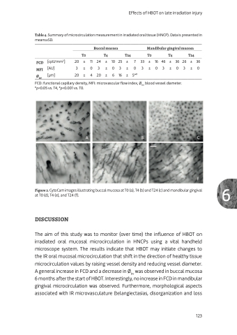
Table 4. Summary of microcirculation measurement in irradiated oral tissue (HNCP). Data is presented in means±SD.
Effects of HBOT on late irradiation injury
         FCD [cpll/mm2] MFI [AU]
[μm]
Buccal mucosa Mandibular gingival mucosa T0 T4 T24 T0 T4 T24
20 ± 11 24 ± 10 25 ± 7 33 ± 16 46 ± 36 26 ± 36 3 ± 0 3 ± 0 3 ± 0 3 ± 0 3 ± 0 3 ± 0
20 ± 4 20 ± 6 16 ± 5*#
ØBV
   FCD: functional capillary density, MFI: microvascular flow index, Øbv: blood vessel diameter. *p<0.05 vs. T4, #p<0.001 vs. T0.
Figure 3. CytoCam images illustrating buccal mucosa at T0 (a), T4 (b) and T24 (c) and mandibular gingival 6 at T0 (d), T4 (e), and T24 (f).
DISCUSSION
The aim of this study was to monitor (over time) the influence of HBOT on irradiated oral mucosal microcirculation in HNCPs using a vital handheld microscope system. The results indicate that HBOT may initiate changes to the IR oral mucosal microcirculation that shift in the direction of healthy tissue microcirculation values by raising vessel density and reducing vessel diameter. A general increase in FCD and a decrease in Øbv was observed in buccal mucosa 6 months after the start of HBOT. Interestingly, no increase in FCD in mandibular gingival microcirculation was observed. Furthermore, morphological aspects associated with IR microvasculature (telangiectasias, disorganization and loss
  123
