
Page 74 - Quantitative Imaging of Small Tumours with Positron Emission Tomography
P. 74
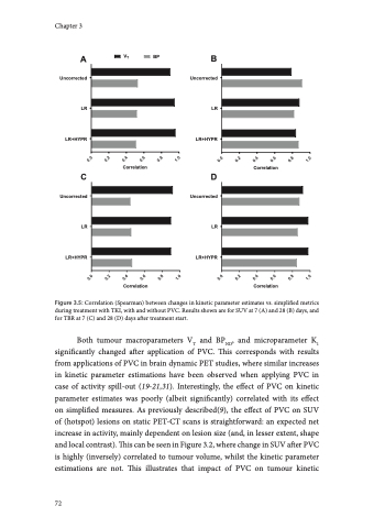
parameter estimation is more complex, as seen in Figure 3.1 which displays the non-linear temporality of partial-volume effects for a typical mediastinal lymph node metastasis. Here, an early spill-in of activity due to blood pool proximity is noted, with increasing activity spill-out afterwards as tumour uptake increases and background activity decreases. Hence, across lesions the effect of PVC on kinetic parameters may differ depending not only on size, but as well on presence of proximate high activity structures, rate of tracer uptake during the scan, and 3 background activity. For quantification of functional tumour characteristic on PET-CT in clinical practice, a simplified quantitative method is necessary, obviating the need for complex and extended dynamic image acquisitions, need for blood sampling, and facilitating the possibility of whole-body acquisitions. To this end, per radiotracer and cancer type simplified metrics needs to be technically validated by pharmacokinetic modeling using dynamic PET-CT (4). In the current study the effect of PVC on kinetic parameter estimates was different from its effect on simplified metrics, which explains why it might affect validation of these simplified metrics (using VT). We observed a trend that PVC increased correspondence of SUV with VT in single measurements (correlations improving from 0.82 to 0.90) and as a biomarker of treatment response (correlations improving from 0.90 to 0.95 at 7 days and from 0.79 to 0.88 at 28 days after treatment start). However, confidence intervals of these correlations overlapped, which might at least partly be due to the sample size (inherent to this type of study), and therefore these differences are not statistically significant. Therefore, while PVC is mandated to acquire accurate quantitative reads, it only increases correspondence of kinetic parameters with simplified metrics to a small extent on a cohort level. This indicates that the impact of image resolution on technical validation of simplified metrics of 18F-FLT as biomarkers of response to TKI might be small, and that PET images without PVC seem non-inferior for this purpose. It should be noted that for response assessment to treatments that affect tracer kinetics and blood pool activity to a larger extent than TKIs and for other cancer types more affected by spill-in (eg. prostate cancer lesions with urinary tract proximity), PVC may have a larger impact on validation of simplified metrics. Spill-out due to PVE will result in overestimation of metabolic tumour volumes, which increases the underestimation of true tracer uptake since background activity is included (11). A parametric PVC method may therefore theoretically reduce inaccuracies in delineation. However, iterative deconvolution PVC in dynamic PET-CT 73
