
Page 65 - Quantitative Imaging of Small Tumours with Positron Emission Tomography
P. 65
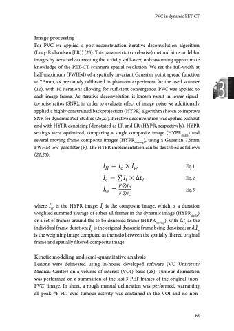
Chapter 3 tumour structures with high uptake were included. Second, this VOI was shrunk to an isocontour based on 50% of the peak value (mean activity in a 12mm sphere positioned to provide the highest uptake value), with correction for local background activity. VOIs were then projected onto each frame of both the original and partial-volume corrected PET images to acquire time activity curves from both the datasets (without and with PVC). To explore the effect of PVC on tumour delineation, tumours were also delineated on the LR+HYPR images using the same approach. Metabolically active tumour volume (MATV) was defined as the sum of voxel volumes within a VOI. A 2x2 voxel (8x8 mm) region was placed centrally in ascending aorta on 5 adjacent slices to acquire an image-derived input function (IDIF), aiming to avoid partial-volume effects. Parent plasma input functions were generated by calibrating IDIFs using the activity concentrations measured in the venous blood samples, and correcting for metabolites and plasma-to-blood ratio. Full quantitative parameters derived from kinetic modeling and simplified measures were extracted using in-house developed software in MATLAB. We used a reversible two-tissue model with blood volume parameter, which has been identified as the optimal compartment model for 18F-FLT by Frings et al. (5). Pharmacokinetic parameters rate of influx of the tracer from blood to tissue (K1), volume of distribution (VT), and binding potential (BPND) of each lesion were derived using non-linear regression, where: Eq.4 Eq.5 VT served as the preferred reference parameter for validation of simplified metrics for 18F-FLT (5). The simplified metrics, mean SUV and tumour-to-blood ratio (TBR; parent plasma), were derived at a 50-60min post-injection scan interval, where: Eq.6 Eq.7 64
