
Page 162 - Quantitative Imaging of Small Tumours with Positron Emission Tomography
P. 162
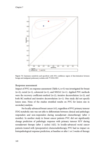
(43). In patients with metastatic colorectal cancer PVC significantly reduced the ROC area-under-the-curve of SUV in discriminating between responders and non-responders after 2 weeks of chemotherapy, as defined with RECIST (44). In NSCLC patients treated with radio- or radiochemotherapy PVC changed PERCIST (3) classification of response in 5/24 lesions, which were verified as correct alterations in clinical follow-up (45). Discussion Quantification of functional tumour characteristics with PET is considered to be useful in clinical oncology, often using semi-quantitative analyses resulting in SUVs. Unfortunately, partial-volume effects are known to cause underestimations of tumour activity, and hence the necessity of PVC for accurate semi-quantitative reads for small lesions is well recognized (5)]. However, many factors affect its accuracy and potentially hamper its optimal usage. Perhaps as a consequence, its resulting advantage in oncological PET studies is not yet evident. Additionally, the lack of consensus on the preferred PVC and delineation method may result in suboptimal results and could hamper comparisons between studies. This review 7 discusses the clinical impact of PVC and gives recommendations for specific research questions and analyses in future studies applying PVC. When applied to diagnosis of primary lesions and (mainly nodal) staging PVC often yielded higher sensitivity at the expense of specificity (Tables 7.1- 7.2 and Figures 7.3-7.4), which is an obvious consequence when using the same test positivity SUV thresholds for uncorrected and PVC data. In the subset of studies which allowed statistical pooling (679 lesions), meta-analysis showed that PVC did not significantly alter the overall diagnostic accuracy of characterizing pulmonary lesions with PET (Figure 7.5). When estimating the effect of PVC, the optimal trade-off between sensitivity and specificity (the SUV threshold of test positivity) may be different for PVC and uncorrected data. At an exploratory level, one should define this cut-off for either method. Of note, Degirmenci et al. (on pulmonary nodules) used data-driven SUV cut-offs of 2.4 and 2.9 for uncorrected and PVC data, respectively, which yielded a specificity fixed at 80% with sensitivities of 62 and 73% for uncorrected and PVC data, respectively (18). We performed a similar analysis using the (individual patient) data from Hickeson et al. (15). At a predefined SUV cutoff of 2.5, PVC decreased specificity Systematic review and meta-analysis 161
