
Page 93 - Fertility in Women with Rheumatoid Arthritis Vruchtbaarheid van vrouwen met reumatoïde artritis
P. 93
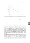
AMH in RA – time to pregnancy
Figure 2 – Kaplan-Meier curve depicting the time to pregnancy in women with rheumatoid arthritis trying to conceive. T=0 is the moment indicated by the patients at which they  rst had actively tried to conceive.
Ninety-four women (45%) were subfertile based on a time to pregnancy >12 months or
a follow-up time >12 months without achieving a pregnancy. For six women (2.9%) it
was unknown whether they ful lled the criteria for subfertility because it was unknown
whether they achieved pregnancy during the follow-up (n=3), or because their follow-up 6 time ended before they had been trying to conceive for 12 months (n=3).
Serum AMH levels and RA disease characteristics
The median preconceptional serum AMH level was 2.5 ug/L (IQR 1.5 – 4.6), with 1 woman having an AMH level below the LOD. The individual preconceptional serum AMH levels were plotted in the nomogram created for healthy controls12 ( gure 3). In 36 women (17.2%) the AMH levels were below the 10th percentile for their age, in 94 patients (45.0%) AMH levels were between the 10th and 50th percentile, in 73 patients (34.9%) between the 50th and 90th percentile, and in 6 (2.9%) of patients AMH levels were above the 90th percentile. ANCOVA with log-transformed AMH levels, showed signi cant lower AMH levels, corrected for age, in patients compared to controls (p=<0.001).
Mono-variable analyses showed a signi cant negative association of log-transformed AMH levels with increasing age (β= -0.070 (95%CI -0.11 ‒ -0.031), p=0.001) and the presence of ACPA (β= -0.38 (95%CI -0.71 ‒ -0.056), p=0.022) (table 2). Serum AMH levels were not associated with never having been pregnant before, disease duration, presence of RF, DAS28-CRP, presence of erosions, use of NSAIDs, use of prednisone,
91
