
Page 138 - Molecular features of low-grade developmental brain tumours
P. 138
5
CHAPTER 5
among the 9400 differentially expressed genes (Table 3). These MMPs and TIMPs were selected for further validation with RT-qPCR. Higher expression of MMP2, MMP11, MMP14, MMP15, MMP19, TIMP1, TIMP2 and TIMP3 was found in SEGA (n=12) as compared to control tissue (n=8; p<0.05; Figure 2a-h). The expression of MMP17 (Figure 2i) was lower in SEGA as
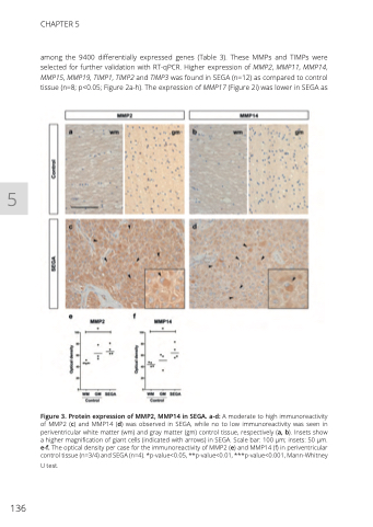
Figure 3. Protein expression of MMP2, MMP14 in SEGA. a-d: A moderate to high immunoreactivity of MMP2 (c) and MMP14 (d) was observed in SEGA, while no to low immunoreactivity was seen in periventricular white matter (wm) and gray matter (gm) control tissue, respectively (a, b). Insets show a higher magnification of giant cells (indicated with arrows) in SEGA. Scale bar: 100 μm; insets: 50 μm. e-f. The optical density per case for the immunoreactivity of MMP2 (e) and MMP14 (f) in periventricular control tissue (n=3/4) and SEGA (n=4). *p-value<0.05, **p-value<0.01, ***p-value<0.001, Mann-Whitney
U test.
136
