
Page 106 - Molecular features of low-grade developmental brain tumours
P. 106
4
CHAPTER 4
104
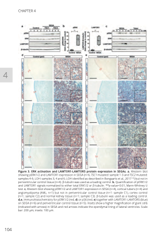
Figure 3. ERK activation and LAMTOR1-LAMTOR5 protein expression in SEGAs. a. Western blot showing pERK1/2 and LAMTOR1 expression in SEGA (n=6; TSC1 mutated: sample 1-3 and TSC2 mutated: samples 4-6; LOH: samples 3, 4 and 6; LOH identified as described in Bongaarts et al., 2017 24) but not in periventricular control tissue (n=4). β-tubulin was used as a loading control. b. Quantification of pERK1/2 and LAMTOR1 signals normalized to either total ERK1/2 or β-tubulin. **p-value<0.01, Mann-Whitney U test. c. Western blot showing pERK1/2 and LAMTOR1 expression in SEGA (n=4), cortical tubers (n=4) and angiomyolipoma (AML; n=1) but not in periventricular control tissue (n=1: sample C1), cortex control (n=1; sample C2) and normal kidney tissue (n=1; sample C3). β-tubulin was used as a loading control. d,e. Immunohistochemistry for pERK1/2 (red, d) or pS6 (red, e) together with LAMTOR1-LAMTOR5 (blue) on SEGA (n=6) and periventricular control tissue (n=5). Insets show a higher magnification of giant cells (indicated with arrows) in SEGA and red arrows indicate the ependymal lining of lateral ventricles. Scale bar: 200 μm; insets: 100 μm.
