
Page 67 - Coronary hemodynamics in acute myocardial infarction - Matthijs Bax
P. 67
The Doppler flow wire in acute myocardial infarction
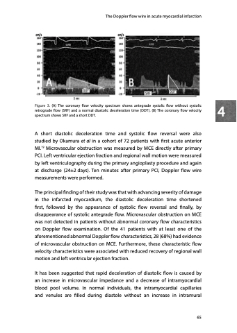
Figure 3. (A) The coronary flow velocity spectrum shows antegrade systolic flow without systolic 4 retrograde flow (SRF) and a normal diastolic deceleration time (DDT). (B) The coronary flow velocity
spectrum shows SRF and a short DDT.
A short diastolic deceleration time and systolic flow reversal were also studied by Okamura et al in a cohort of 72 patients with first acute anterior MI.19 Microvascular obstruction was measured by MCE directly after primary PCI. Left ventricular ejection fraction and regional wall motion were measured by left ventriculography during the primary angioplasty procedure and again at discharge (24±2 days). Ten minutes after primary PCI, Doppler flow wire measurements were performed.
The principal finding of their study was that with advancing severity of damage in the infarcted myocardium, the diastolic deceleration time shortened first, followed by the appearance of systolic flow reversal and finally, by disappearance of systolic antegrade flow. Microvascular obstruction on MCE was not detected in patients without abnormal coronary flow characteristics on Doppler flow examination. Of the 41 patients with at least one of the aforementioned abnormal Doppler flow characteristics, 28 (68%) had evidence of microvascular obstruction on MCE. Furthermore, these characteristic flow velocity characteristics were associated with reduced recovery of regional wall motion and left ventricular ejection fraction.
It has been suggested that rapid deceleration of diastolic flow is caused by an increase in microvascular impedance and a decrease of intramyocardial blood pool volume. In normal individuals, the intramyocardial capillaries and venules are filled during diastole without an increase in intramural
65
