
Page 66 - Coronary hemodynamics in acute myocardial infarction - Matthijs Bax
P. 66
Chapter 4
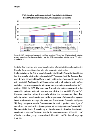
Figure 2. CFVR, Baseline and hyperaemic peak flow velocity in IRAs and non-IRAs immediately after the primary procedure, after 1 week and after 6 months. CFVR, coronary flow velocity reserve; IRA, infarct- related artery.
Systolic flow reversal and rapid deceleration of diastolic flow: characteristic Doppler flow-velocity patterns in microvascular obstruction
Iwakura et al were the first to report characteristic Doppler flow velocity patterns in microvascular obstruction after acute MI.11 They examined the Doppler-flow wire derived coronary blood flow velocity pattern in 42 consecutive patients with acute MI. Additionally, MCE was performed in all patients both before and after primary angioplasty. Microvascular obstruction was detected in 11 patients (26%) by MCE. The coronary flow velocity pattern appeared to be normal in patients without microvascular obstruction on MCE (Figure 3a). However, in patients with microvascular obstruction the coronary blood flow velocity pattern was characterized by the appearance of abnormal retrograde flow in early systole, and rapid deceleration of the diastolic flow velocity (Figure 3b). Early retrograde systolic flow was seen in 10 of 11 patients with signs of no reflow compared with only one patient without signs of no reflow on MCE. The rate of decline in flow velocity in diastole was calculated as the diastolic deceleration rate (cm/s2). Mean diastolic deceleration rate was 106.4±76.1 cm/ s2 in the no-reflow group compared with 55.9±31.2 cm/s2 in the reflow group (p<0.01).
64
