
Page 126 - Coronary hemodynamics in acute myocardial infarction - Matthijs Bax
P. 126
Chapter 7
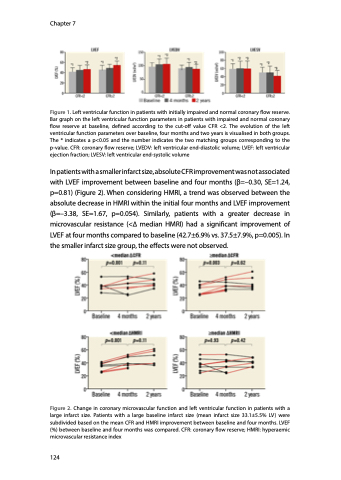
Figure 1. Left ventricular function in patients with initially impaired and normal coronary flow reserve. Bar graph on the left ventricular function parameters in patients with impaired and normal coronary flow reserve at baseline, defined according to the cut-off value CFR <2. The evolution of the left ventricular function parameters over baseline, four months and two years is visualised in both groups. The * indicates a p<0.05 and the number indicates the two matching groups corresponding to the p-value. CFR: coronary flow reserve; LVEDV: left ventricular end-diastolic volume; LVEF: left ventricular ejection fraction; LVESV: left ventricular end-systolic volume
In patients with a smaller infarct size, absolute CFR improvement was not associated with LVEF improvement between baseline and four months (β=–0.30, SE=1.24, p=0.81) (Figure 2). When considering HMRI, a trend was observed between the absolute decrease in HMRI within the initial four months and LVEF improvement (β=–3.38, SE=1.67, p=0.054). Similarly, patients with a greater decrease in microvascular resistance (<∆ median HMRI) had a significant improvement of LVEF at four months compared to baseline (42.7±6.9% vs. 37.5±7.9%, p=0.005). In the smaller infarct size group, the effects were not observed.
Figure 2. Change in coronary microvascular function and left ventricular function in patients with a large infarct size. Patients with a large baseline infarct size (mean infarct size 33.1±5.5% LV) were subdivided based on the mean CFR and HMRI improvement between baseline and four months. LVEF (%) between baseline and four months was compared. CFR: coronary flow reserve; HMRI: hyperaemic microvascular resistance index
124
