
Page 120 - Helicobacter pylori and Gastric Cancer: From Tumor microenvironment to Immunotherapy
P. 120
114
Incomplete 30.0% (12/40) 31.6% (12/38) <0.001 33.5 (7.4–37.3)
Unidentified 42.5 % (17/40) 36.84(14/38) <0.001 41.7 (4.1–74.1) Chapter 5
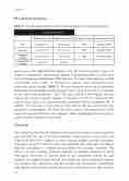
IM, intestinal metaplasia
Chapter 5
Table 8. The association between OLGA gastritis stage and intestinal metaplasia
OLGA gastritis stage
Stage 0–II (n = 121)
Stage III, IV (n = 47)
p (Fisher’s exact)
Odd ratios (95% CI)
IM
49.58 (60/121)
38.29 (18/47)
<.001
24.6 (05.7–72.5)
113
Extensive
26.45 (32/121)
25.53 (12/47)
<.001
22.6 (07.8–82.9)
Incomplete subtype
18.18 (22/121)
21.27 (10/47)
<.001
33.7 (04.5–62.8)
Unidentified subtype
4.13 (7/121)
0 (0/47)
Seven cases with unidentified IM subtype were all in OLGA gastritis stage 0–II. When we subsequently analyzed only patients with gastritis stage 0–II only, there was no homogenous distribution of IM subgroup: We rarely found patients with IM none-to-mild EGA, while it clustered to patients with moderate-to-severe endoscopic gastric atrophy (Table 7). We also found that there was an association between the gastric atrophy and the subtype of IM. From a total of 59.7% (148 /248) in our entire study population, 46(37.1%) and 32(25.8%) from Nigeria and Iran respectively had the complete subtype of IM while 30 (24.2%) and 26 (20.9%) patients from Nigeria and Iran respectively presented with the incomplete IM. In addition, 9 (7.2%) and 5 (4.1%) patients from Nigeria and Iran respectively were classified as indeterminate IM. Thus also with respect to the presentation of IM, important regional differences are apparent, further highlighting the necessity to tailor screening strategies to local needs.
Discussion.
The natural history of the development and progression of gastric cancer in general and especially the role of H. pylori infection in this process is now fairly well understood and involves sequence of gastric mucosal atrophy, intestinal metaplasia, and gastric cancer[23]. This has led to the realization that endoscopic screening in high-risk individuals is essential for preventing the associated mortality. The efficacy of such screening obviously depends on adequate diagnosis in which histological assessment of biopsies taken by the endoscopist remains the gold standard. The updated Sydney system was designed to assess histological gastritis and atrophy more objectively and has become the international standard[24]. Although this classification includes assessment of five biopsy sites, this extensive
118
