
Page 41 - Towards personalized therapy for metastatic prostate cancer: technical validation of [18F]fluoromethylcholine
P. 41
Dual-phase [18F]FCH PET/CT in prostate cancer
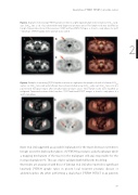
Figure 3. Example of decreasing [18F]FCH uptake over time in a right inguinal lymph node (red arrow; SUVmax early: 4.41; SUVmax late: 2.03) of a patient with newly diagnosed prostate cancer. This lymph node was classified as benign. Transversal sections of the Low-dose CT, PET and fused PET/CT images: a, b and c – early phase; d, e and f – late phase. [18F]FCH uptake in the prostate is also visible.
2
Figure 4. Example of increasing [18F]FCH uptake over time in a right para-iliac lymph node (red solid arrow; SUVmax early: 3.02; SUVmax late: 4.35) and a left para-iliac node (red dotted arrow; SUVmax early: 3.47; SUVmax late: 4.79) of a patient with biological relapse after initially treated prostate cancer. These lymph nodes were classified as malignant. Transversal sections of the Low-dose CT, PET and fused PET/CT images: a, b and c – early phase; d, e and f – late phase.
Kwee et al. [15] suggested as a possible explanation for the tracer decrease over time in benign zones the dephosphorylation of [18F]FCH by prostatic acid phosphatase while a trapping mechanism of the tracer in the malignant cells was responsible for the increased uptake in PC. This can only be validated with full kinetic modeling.
Our results are at variance with those of Cimitan et al. [15] who reported no significant late/early [18F]FCH uptake ratios in proven local recurrent prostatic disease or abdominopelvic LN, when performing a dual phase [18F]FCH PET/CT in 43 patients
39
