
Page 45 - Development of Functional Scaffolds for Bone Tissue Engineering Using 3D-Bioprinting of Cells and Biomaterials - Yasaman Zamani
P. 45
efficiency of scaffold groups was similar; (iii) proliferation and collagenous matrix deposition was highest in the RGD-immobilized scaffolds; (iv) ALP activity was higher in NaOH-treated scaffolds compared to RGD-immobilized scaffolds; (v) NaOH-treated scaffolds, but not RGD-immobilized scaffolds, increased calcium deposition. Therefore, our results showed enhanced osteogenic differentiation potential in 24 h NaOH-treated scaffolds compared to RGD-immobilized scaffolds in vitro, suggesting that chemical treatment of 3D-printed PCL scaffolds by 3 M NaOH for 24 h might be more promising for in vivo bone regeneration than RGD immobilization.
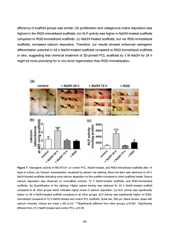
Figure 7. Osteogenic activity of MC3T3-E1 on control PCL, NaOH-treated, and RGD-immobilized scaffolds after 14 days of culture. (a) Calcium mineralization visualized by alizarin red staining. More red stain was observed on 24 h NaOH-treated scaffolds indicating more calcium deposition on this scaffold compared to other scaffolds tested. Scarce calcium deposition was observed on unmodified controls, 72 h NaOH-treated scaffolds, and RGD-immobilized scaffolds. (b) Quantification of the staining. Higher optical density was obtained for 24 h NaOH-treated scaffold compared to all other groups which indicates higher levels of calcium deposition. (c) ALP activity was significantly higher on 24 h NaOH-treated scaffold compared to all other groups. ALP activity was significantly higher on RGD- immobilized compared to 72 h NaOH-treated and control PCL scaffolds. Scale bar, 500 μm. Black arrows, areas with calcium minerals. Values are mean ± SD (n=3). ***Significantly different from other groups, p<0.001, #significantly different from 72 h NaOH-treated and control PCL, p<0.05.
43
