
Page 14 - Wondergem
P. 14
Chapter 1
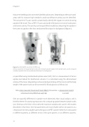
that are emitted by positron emitter labelled radiotracers. Depending on the tracer used, areas with for instance high metabolic and/or proliferative activity can be identified. The concurrent CT scan is used to anatomically identify the organs or tumors showing uptake of the tracer. Thus, a PET-CT scan can provide information on tumor localization and tumor activity. This activity can be quantified visually by comparing the uptake in the tumor to uptake in the liver, mediastinal blood pool or background (figure 1),
Figure 1 A: 18F-FDG PET scan. B 18F-FLT PET scan
Arrows: examples of enlarged lymph nodes with increased uptake. Uptake in the lymph nodes can be compared visually to uptake in the liver, mediastinal blood pool, or background.
or quantified using standardised uptake value (SUV). SUV is a measurement of tumor uptake normalised for distribution volume. It is calculated using the administered activity of the tracer (depending on dose and physical decay of the tracer) and the body weight of the patient and can be corrected for blood glucose as follows (21):
SUV= activity measured in the volume of interest (kBq/ml) , for correction: x glucose patient (mmol/l) administered activity (MBq)/ body weight (kg) 5.0 mmol/l
SUV can quantify differences in uptake more objectively than visual analysis, and is therefore better for assessing response or for comparing uptake between lymph nodes (22). SUVmax is the SUV in the voxel with maximum uptake and is used in the studies described in this thesis. SUV measurements are most reliable within one patient, at a certain point in time using the same settings on one PET scanner. When SUV is measured in different patients, at different times in the same patient, or on different scanners,
12
