
Page 69 - Recognizing axial spondyloarthritis - Janneke de Winter
P. 69
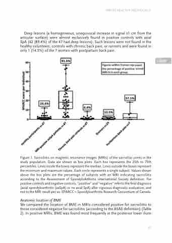
Deep lesions (a homogeneous, unequivocal increase in signal ≥1 cm from the articular surface) were almost exclusively found in positive controls with axial SpA (42 (89.4%) of the 47 had deep lesions). Such lesions were not found in the healthy volunteers, controls with chronic back pain, or runners and were found in only 1 (14.3%) of the 7 women with postpartum back pain.
Figure 1. Sacroiliitis on magnetic resonance images (MRIs) of the sacroiliac joints in the study population. Data are shown as box plots. Each box represents the 25th to 75th percentiles. Lines inside the boxes represent the median. Lines outside the boxes represent the minimum and maximum values. Each circle represents a single subject. Values shown above the box plots are the percentage of subjects with an MRI indicating sacroiliitis according to the Assessment of SpondyloArthritis international Society definition. For positive controls and negative controls, “positive” and “negative” refer to the final diagnosis (axial spondyloarthritis (axSpA) or no axial SpA) after vigorous diagnostic evaluation, and not to the MRI result per se. SPARCC = Spondyloarthritis Research Consortium of Canada.
Anatomic location of BME
We compared the location of BME in MRIs considered positive for sacroiliitis to those considered negative for sacroiliitis (according to the ASAS definition) (Table 2). In positive MRIs, BME was found most frequently in the posterior lower ilium
MRI IN HEALTHY INDIVIDUALS
                                                                 67
 FOUR
