
Page 118 - Recognizing axial spondyloarthritis - Janneke de Winter
P. 118
CHAPTER SEVEN
criteria. However, inflammatory lesions on MRI (7 (41%) versus 3 (9%); P=0.007) and radiographic signs of sacroiliitis (3 (18%) versus 0 (0%); P=0.014) were found more often in those who fulfilled the ASAS axial SpA and/or ESSG classification criteria. In conclusion, first-degree relatives who fulfilled the ASAS axial SpA and/ or ESSG classification criteria had more axial, entheseal, and joint pain, which was reflected in higher disease activity and worse function, than those who did not. Except for more frequent bone marrow edema on MRI, however, they did not show objective signs of inflammation such as clinical arthritis or elevated levels of acute-phase reactants.
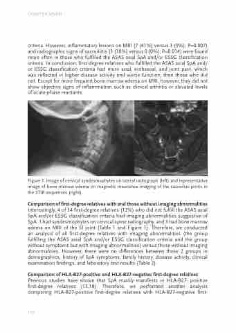
Figure 1. Image of cervical syndesmophytes on lateral radiograph (left) and representative image of bone marrow edema on magnetic resonance imaging of the sacroiliac joints in the STIR sequences (right).
Comparison of first-degree relatives with and those without imaging abnormalities
Interestingly, 4 of 34 first-degree relatives (12%) who did not fulfill the ASAS axial SpA and/or ESSG classification criteria had imaging abnormalities suggestive of SpA: 1 had syndesmophytes on cervical spine radiography, and 3 had bone marrow edema on MRI of the SI joint (Table 1 and Figure 1). Therefore, we conducted an analysis of all first-degree relatives with imaging abnormalities (the group fulfilling the ASAS axial SpA and/or ESSG classification criteria and the group without symptoms but with imaging abnormalities) versus those without imaging abnormalities. However, there were no differences between these 2 groups in demographics, history of SpA symptoms, family history, disease activity, clinical examination findings, and laboratory test results (Table 2).
Comparison of HLA-B27-positive and HLA-B27-negative first-degree relatives
Previous studies have shown that SpA mainly manifests in HLA-B27- positive first-degree relatives (13,18). Therefore, we performed another analysis comparing HLA-B27-positive first-degree relatives with HLA-B27-negative first-
116
