
Page 19 - Clinical relevance of current materials for cranial implants
P. 19
General introduction and outline of this thesis
Materials used for cranioplasties 1 Autologous bone
Von Walther performed the first human cranioplasty with autologous bone in 1821.
Many other surgeons followed him: Ollier(1859), who believed that the periosteum was
the most important tissue for bone regeneration. William MacEwen (1878) successfully inserted fractured calvarial bone and reinserted bone after trepanation. Seydel (1889) used tibial autografts for cranial repair, Muller (1890) developed the “sliding flaps” technique of the external tabula, and Beck (1906) introduced temporal muscle and fascia for the reconstruction of cranial defects. Dobrothworski (1911) used whole ribs, Röpke (1912) scapula, Mauclaire (1914) ilium, and split ribs were described by Brown (1917), all for the repair of cranial defects17,18.
Nowadays autologous bone is still used for cranioplasties. Autologous bone does not suffer from immune rejection, and bony ingrowth and revascularization have been observed14,19. However, it is associated with a high risk of complications.
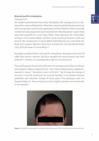
The most frequently mentioned complications for autologous bone flaps are infection and resorption. Infection ranges from 0% - 30%14 and is mostly caused by Staphylococci, especially S. Aureus20. Resorption occurs in 0%-50%19. Up till today the etiology of resorption is not fully understood. We do know that there is an imbalance between osteoblasts and osteoclasts, because of which parts of the autologous bone will disappear (Figure 3). These complications lead to high re-operation and removal rates of cranioplasties20.
Figure 3: A patient who underwent a cranioplasty of autologous bone; after 18 months resorption was observed.
17
