
Page 134 - Clinical relevance of current materials for cranial implants
P. 134
132
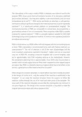
Chapter 6
The cranioplasty in this study is made of PEEK, a relatively new material used for this purpose. PEEK shows good chemical resistance because of its resonance-stabilized and aromatic structure10, has long term stability in wet environments, and can resist temperatures up to 260°C11,12. PEEK can be sterilizedin an autoclave or with gamma- sterilization without significant changes to the material properties; it can be repeatedly sterilized13,14. It is radiolucent without artefacts on (postoperative) imaging15. The mechanical properties of PEEK are comparable to cortical bone; biocompatibility is good without release of ions or constituents. These properties make PEEK a suitable material for medical implants15,16. PEEK is a versatile material, suitable for CAD-CAM technology using a direct production method: no mold or intra-operative production procedures are necessary17.
PEEK is not bioactive, so a PEEK surface will not integrate with the surrounding tissues as bone. PEEK cranioplasty is recommended to be used with fixation material, e.g. osteosynthesis15,16. The risk of infection is one of the main disadvantages and the most important complication reported in literature18. Higher costs are an important issue too. A PEEK PSI, including a resection template and a control template, adds up to approximately 7500 EUR including work-up in the Netherlands. However, the preoperative planning time is approximately 1 hour. With only one procedure is needed, total cost and surgical time are likely lower compared to a two-staged surgical procedure. Raw PEEK is a relatively expensive material which has to be milled; in this process, a great portion of the material becomes unusable19.
Other designs for resection templates in cranial defects have been recently described. In the design of Carolus et al., only the outline of the resection is established in the template20. In our study the resection template forces the surgeon to follow the resection outline through the use of an inner and outer piece of the template. The inner part of the resection template ensures that the meningioma can removed in one piece (figure 4C). The design of the resection template is important to make the surgical intervention easier and reduce operation time.
