
Page 131 - Clinical relevance of current materials for cranial implants
P. 131
Resection templates with 3D virtual planning
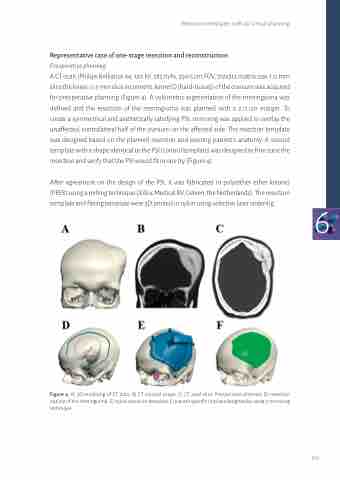
Representative case of one-stage resection and reconstruction
Preoperative planning
A CT-scan (Philips Brilliance 64, 120 kV, 285 mAs, 25x15 cm FOV, 512x512 matrix size, 1.0 mm slice thickness, 0.5 mm slice increment, kernel D (hard-tissue)) of the cranium was acquired for preoperative planning (Figure 4). A volumetric segmentation of the meningioma was defined and the resection of the meningioma was planned with a 2.0 cm margin. To create a symmetrical and aesthetically satisfying PSI, mirroring was applied to overlay the unaffected, contralateral half of the cranium on the affected side. The resection template was designed based on the planned resection and existing patient’s anatomy. A second template with a shape identical to the PSI (control template) was designed to fine-tune the resection and verify that the PSI would fit in one try (Figure 4).
After agreement on the design of the PSI, it was fabricated in poly(ether ether ketone)
(PEEK) using a milling technique (Xilloc Medical BV, Geleen, the Netherlands). The resection
template and fitting template were 3D printed in nylon using selective laser sintering. 6
Figure 4: A) 3D rendering of CT data, B) CT coronal coupe, C) CT axial slice. Preoperative planned, D) resection outline of the meningioma, E) nylon resection template, F) patient specific implant designed by using a mirroring technique
129
