
Page 163 - Fluorescence-guided cancer surgery
P. 163
Clinical translation of OTL38 in ovarian cancer 161
form as being either uorescent or non- uorescent and as being either clinically suspected of malignancy or not (Figure S1, online available). All resected lesions were examined for tumor status by an experienced pathologist. A positive tumor that was uorescent was considered a true positive; a negative lesion that was uorescent was considered a false positive; and a positive tumor that was non- uorescent was considered a false negative. In addition, we performed immunohistochemistry to demonstrate FRα and FRβ expression coupled with uorescence microscopy in order to evaluate OTL38 binding (Supplementary Materials and Methods, online available).
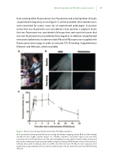
Figure 1. Fluorescence measurements in the skin of healthy volunteers
A. A researcher measuring skin uorescence using the Artemis imaging system. B. Color (left column) and uorescence (right column) images of a healthy volunteer at baseline (upper row) and after receiving a dose of OTL38 (lower row). Note that a bright uorescent signal is detected after dosing, and uorescence intensity can be measured within a region of interest (ROI) (dashed circle). C. Fluorescence intensity (measured in arbitrary units) in an ROI over time (in hours). The uorescence signal reached peak levels at approximately 2 hours and was stable for up to 6 hours, then decreased over the following 48 hours.
