
Page 14 - Fluorescence-guided cancer surgery
P. 14
12
Chapter 1
structures10. This technique, like medical imaging techniques in general, is based on the ability to create a contrast ratio between the tissue of interest and its surrounding normal tissue.
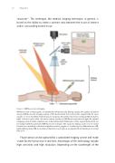
Figure 1. NIR uorescence imaging
NIR uorescent contrast agents are administered intravenously. During surgery, the agent is visualized using a NIR uorescent imaging system of the desired form factor (above the surgical eld for open surgery or encased within minimal invasive surgery). All systems must have adequate NIR excitation light, collection optics, lter sets and a camera sensitive to NIR uorescent emission light. An optimal imaging system includes simultaneous visible (white) light illumination of the surgical eld, which can be merged with the generated NIR uorescence images. The surgeon’s display can be one of several form factors, including a standard computer monitor, goggles or a wall projector. Abbreviations: LED, light emitting diode; NIR, near-infrared. Illustration and caption are depicted from Vahrmeijer et al., Nat Rev 201310.
Fluorescence can be captured by a specialized imaging system and made visible for the human eye in real-time. Advantages of this technology include high sensitivity and high resolution. Depending on the wavelength of the
