
Page 54 - Clinical variability in Noonan syndrome with emphasison ear and eye
P. 54
CHAPTER 3
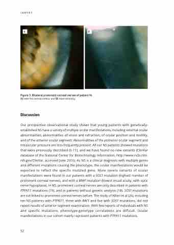
Figure 3. Bilateral prominent corneal nerves of patient 14. (A) near the corneal limbus, and (B) more centrally.
Discussion
Our prospective observational study shows that young patients with genetically- established NS have a variety of multiple ocular manifestations, including external ocular abnormalities, abnormalities of vision and refraction, of ocular position and motility, and of the anterior ocular segment. Abnormalities of the posterior ocular segment and intraocular pressure are less-frequently present. All our NS patients showed mutations that were previously described (6-11), and we have found no new variants (ClinVar database of the National Center for Biotechnology Information, http://www.ncbi.nlm. nih.gov/ClinVar, accessed June 2016). As NS is a clinical diagnosis with multiple genes and different mutations causing the phenotype, the ocular manifestations would be expected to reflect the specific mutated gene. More severe variants of ocular manifestations were found in our patients with a SOS1 mutation (highest number of prominent corneal nerves), and with a BRAF mutation (lowest visual acuity, with optic nerve hypoplasia). In NS, prominent corneal nerves are only described in patients with PTPN11 mutations (19), and in patients without genetic analysis (18). SOS1 mutations are not linked to prominent corneal nerves before. The study of Alfieri et al (26), including ten NS patients with PTPN11, three with RAF1 and five with SOS1 mutations, did not report results of anterior segment examination. With few reports of individuals with NS and specific mutations, phenotype-genotype correlations are difficult. Ocular manifestations in our cohort mainly represent patients with PTPN11 mutations.
52
