
Page 18 - Sentinel lymph node biopsy in oral cavity cancer - Inne J. den Toom
P. 18
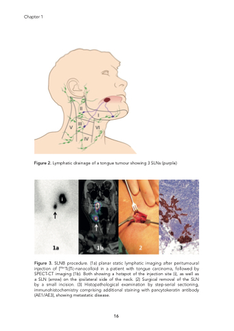
Chapter 1 Figure 2. Lymphatic drainage of a tongue tumour showing 3 SLNs (purple) Figure 3. SLNB procedure. (1a) planar static lymphatic imaging after peritumoural injection of [99mTc]Tc-nanocolloid in a patient with tongue carcinoma, followed by SPECT-CT imaging (1b). Both showing a hotspot of the injection site (i), as well as a SLN (arrow) on the ipsilateral side of the neck. (2) Surgical removal of the SLN by a small incision. (3) Histopathological examination by step-serial sectioning, immunohistochemistry comprising additional staining with pancytokeratin antibody (AE1/AE3), showing metastatic disease. 16
