
Page 119 - Assessing right ventricular function and the pulmonary circulation in pulmonary hypertension Onno Anthonius Spruijt
P. 119
simultaneously; enabling advanced hemodynamic characterization of individual patients in clinical practice [61].
A limitation of the combined measurement of pressure and MRI derived flow is that a right sided heart catheterization is still required and that for simultaneous measurements an interventional (X- )MRI suite together with a special catheterization set must be used. Thus, although this approach can be of use in conditions that an accurate invasive stroke volume measurement is not possible, or in experimental settings, it is unlikely that this type of measurement will be performed routinely in the near future.
MRI pulmonary angiography and perfusion measurements
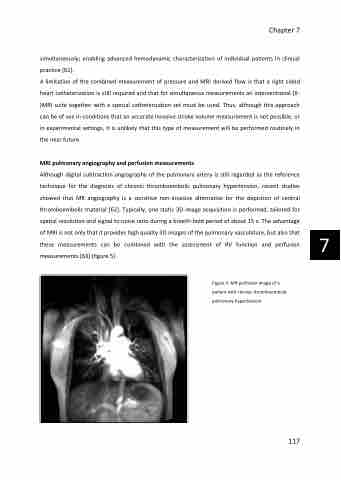
Although digital subtraction angiography of the pulmonary artery is still regarded as the reference technique for the diagnosis of chronic thromboembolic pulmonary hypertension, recent studies showed that MR angiography is a sensitive non-invasive alternative for the depiction of central thromboembolic material [62]. Typically, one static 3D image acquisition is performed, tailored for spatial resolution and signal to noise ratio during a breath-hold period of about 15 s. The advantage of MRI is not only that it provides high quality 3D images of the pulmonary vasculature, but also that these measurements can be combined with the assessment of RV function and perfusion measurements [63] (figure 5).
Chapter 7
Figure 5: MR perfusion image of a patient with chronic thromboembolic pulmonary hypertension
117
7
