
Page 64 - Tailoring Electrospinning Techniques for Regenerative Medicine - Marc Simonet
P. 64
CHAPTER 3
3.4 Results and discussion
3.4.1 Mesh preparation
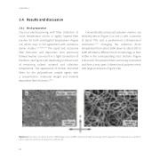
Classical electrospinning with fiber collection at room temperature results in tightly layered fiber meshes for both investigated biopolymers (Figure 3.2) which stays in full agreement with numerous earlier studies.2,11,17,18,36,37 The rapid and successive fiber formation and deposition onto previously formed meshes can result in a tight connection of the fibers (see Figure 3.2b) depending on the amount of remaining solvent, material and collection temperature. The appearance of thicker, branched fibers for the polyurethane sample agrees with a concentration, molecular weight and material dependent fiber thickness.38,39
Conventionally processed polymer meshes are relatively dense (Figure 3.3a and c) with a porosity of about 79% and a predominant 2-dimensional orientation.40,41 Changing the collection drum temperature from about 300K down to about 200 to 220K a orded a di erent mesh morphology as best visible in the corresponding cross sections (Figure 3.3b and d). The polymer fibers are loosely connected and form a very open 3-dimensional polymer mesh with large void spaces (Figure 3.3b).
Figure 3.2 Top view on polymer meshes (SEM images) spun at 30% relative humidity and using a drum target at room temperature; poly(lactic acid-co-glycolic acid) (a) and polyester-urethane (b).
62
