
Page 85 - scheppingen
P. 85
DYSREGULATION OF THE (IMMUNO)PROTEASOME PATHWAY IN MCD
hours (same fixation time used for the surgical specimens); no differences in the immu- noreactivity pattern were observed.
Single-label immunohistochemistry was performed as previously described 25. Sections were deparaffinated in xylene, rinsed in ethanol (100%, 95%, 70%) and incu- bated for 20 minutes in 0.3% hydrogen peroxide diluted in methanol. Antigen retrieval was performed using a pressure cooker in 0.1 M citrate buffer pH 6.0 at 120°C for 10 minutes. Slides were washed with phosphate-buffered saline (PBS; pH 7.4) and incu- bated overnight with the primary antibody in PBS at 4°C. After washing in PBS, sec- tions were stained with a polymer based peroxidase immunohistochemistry detection kit (PowerVision Peroxidase system, ImmunoVision, Brisbane, CA, USA). The 3,3’-diami- nobenzidine tetrahydrochloride was used as chromogen. Sections were dehydrated in alcohol and xylene and coverslipped.
Double-labeling of β1, β1i, β5 or β5i with NeuN (neuronal nuclear protein (NeuN; mouse clone MAB377; Chemicon, Temecula, CA, USA; 1:2000), GFAP (polyclonal rabbit, DAKO, Glostrup, Denmark, 1:4000; or monoclonal mouse, Sigma, St. Louis, Mo, USA; 1:4000), HLA-I (mouse clone HC-10, 1:200) or HLA-II (mouse anti-human leuko- cyte antigen (HLA)-DP, DQ, DR, mouse clone CR3/43; DAKO; 1:400) was performed as previously described 26). Sections were incubated with Brightvision poly-alkaline phos- phatase (AP)-anti-rabbit or anti-mouse (Immunologic, Duiven, The Netherlands) for 30 minutes at room temperature, and washed with PBS. AP activity was visualized with the AP substrate kit III Vector Blue (SK-5300, Vector laboratories Inc., CA, USA). To remove the first primary antibody sections were incubated at 121 °C in citrate buffer (10 mM NaCi, pH 6.0) for 10 min. Incubation with the second primary antibody was performed
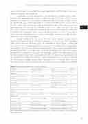
Table 2 Immunochistochemistry: primary antibodies
Antigen Primary Antibody Source Dilution
Glial fibrillary acidic protein Rabbit polyclonal DAKO, Glostrup, Denmark 1:4000 (GFAP)
Neuronal nuclear protein Mouse clone MAB377 Chemicon, Temecula, CA, 1:2000 (NeuN) USA
Phospho-S6 ribosomal pro- Ser235/236; rabbit polyclonal Cell Signaling Technology, 1:50 tein (pS6) Beverly, MA, USA
Interleukin 1β Goat polyclonal Santa Cruz Bio., Delaware 1:70 CA, USA
MHC class I (HLA A, B and C; Mouse clone HC-10 * 1:200 HLA-I)
MHC class II (HLA-DP, DQ, Mouse clone CR3/43 DAKO, Glostrup, Denmark 1:400 DR; HLA-II)
Proteasome β1 Mouse monoclonal IgG1 Enzo Life Sciences/Biomol, 1:200 Farmingdale, NY, USA
Proteasome β5 Rabbit polyclonal Enzo Life Sciences/Biomol 1:500
Proteasome β1i Mouse monoclonal IgG1 Enzo Life Sciences/Biomol 1:200
Proteasome β5i Mouse monoclonal IgG1 Enzo Life Sciences/Biomol 1:200
MHC: major histocompatibility complex; * gift from Prof. J. Neefjes, Netherlands Cancer Institute, The Netherlands.
83
four
