
Page 129 - scheppingen
P. 129
EXPRESSION OF MICRORNAS MIR21, MIR146A, AND MIR155 IN TSC
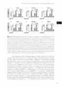
Figure 6 miRNA expression levels after modulation in cultures. Quantitative real-time PCR of miR21 (A and D), miR146a (B and E) and miR155 (C and F) expression in human fetal astrocytes and SEGA-derived cell cultures after transfection with either mimic or inhibitor for the corresponding miRNA. Cultures were transfected during 24 hours, followed by 24 hours exposure to IL-1β (10 ng/ml). Transfection with miR21, miR146a and miR155 mimic increased the expression levels of the corresponding miRNAs substantially compared to control, both in cells exposed to IL-1β (10 ng/ ml) and in unstimulated cells. Transfection with miR21, miR146a or miR155 inhibitors 24 hours be- fore IL-1β exposure prevented the IL-1β-induced increased expression of the three miRNAs. Data are expressed relative to the levels observed in untreated cells and are mean ± SEM from five fetal and four SEGA experiments on cultures derived from separate donors performed in triplicate. *p<0.05, **p<0.01, ***p<0.001 compared to control; ##p<0.01, ###p<0.001 between experimental samples, Mann Whitney U test. SEGA: subependymal giant cell astrocytomas.
In the present study we provide evidence of both induction and release of miR146a upon stimulation with IL-1β in both cell types. Extracellular release of miRNAs has been shown to take place through different processes, including passive leakage from dying cells, as well as active secretion via microvesicles or using a microvesicle-free pathway 71, 72. However, under our experimental conditions cell viability was not influ- enced by IL-1β stimulation or treatment with miRNA mimic, making passive release from dying cells less likely. Extracellular miRNAs, may represent a new form of intercellu- lar communication, acting as signaling molecules 72-74 and a promising class of biomarkers in different neurological conditions, including epilepsy 75. Interestingly, increased miR146a plasma levels have been reported in a rat model of TLE 34, as well as in a large cohort of epilepsy patients compared with controls 76. Emerging evidence indicates that the level of miR146a may influence seizure activity in experimental models, through modulation of the inflammatory response (77, 78; unpublished observation). Accordingly, miRNA146a has been identified as key negative-feedback regulator of the astrocyte-mediated inflam- matory response (for reviews see 27, 28, 33. In the present study, we confirm the ability of
127
five
