
Page 125 - scheppingen
P. 125
EXPRESSION OF MICRORNAS MIR21, MIR146A, AND MIR155 IN TSC
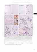
Figure 4 In situ hybridization of miR155 in control, perituberal cortex and TSC cortical tuber and SEGA. A-B: miR155, control grey (A) and white matter (B); miR155 was expressed in neurons (ar- rows in A), but was undetectable in glial cells. C: perituberal cortex (peri-ctx), showing low expres- sion of miR155. D-H (cortical tuber), showing expression of miR55 within the tuber; arrows in D indicate dysmorphic neurons, arrowheads point to a positive blood vessel, asterisks in D indicates positive giant cells. Inserts in E show miR155 expression in GFAP (a), NeuN (b) and CD34 positive cells (c; endothelial cells); arrows in F indicate giant cells, arrowheads point to a positive blood vessel; arrows in D and H indicate dysmorphic neurons; arrowheads in G indicate a positive blood vessel; arrowheads in H indicate positive glial cells. I: miR155 expression in subependymal giant cell astrocytoma (SEGA). Scale bar in I: A-E: 80 μm. F-I: 40 μm.
123
five
