
Page 91 - Imaging of Osteoarthritis and Rheumatoid Arthritis in Hand Joints
P. 91
High-resolution MRI of cartilage in finger joints
In the hand OA patients, normal inter-bone distance was found in 110/161 (68%) joints (41 PIP and 69 MCP joints). In these joints, 33 (24 PIP and 9 MCP) showed thinning of the cartilage layer on the SPGR images of which 14 (12 PIP and 2 MCP) showed areas with full thickness cartilage loss (see Table 4 and Figure 2 for an example). In total seven (3 PIP and 4 MCP) joints in the hand OA patients showed no cartilage damage on SPGR images, but they were scored as abnormal using the inter bone distance with the coronal PD images. In the healthy controls, reduced inter-bone distance was found in 11 (10 PIP and 1 MCP) joints, of which 9 did not show cartilage loss on the SPGR images. Using the hrMRI cartilage score, the readers scored 2 PIP joints in healthy controls as abnormal.
Table 4. Number of joints with cartilage damage split by joint type (n=232)
joints in HOA patients
joints in healthy controls
PIP joints n=81
38
60
Full thickness cartilage loss on 31 6 22 0 0 0 5 hrMRI
Inter bone distance >0 hrMRI cartilage score >0
MCP joints n=80
12
17
Total pat. n=41
23
34
PIP joints n=36
10
2
MCP joints n=35
1
0
Total hc n=18
7
2
hrMRI = high resolution MRI; hc = healthy controls; MCP = metacarpal phalangeal joint; DIP = distal interphalangeal joint. Presented values are means of the two readers (rounded down).
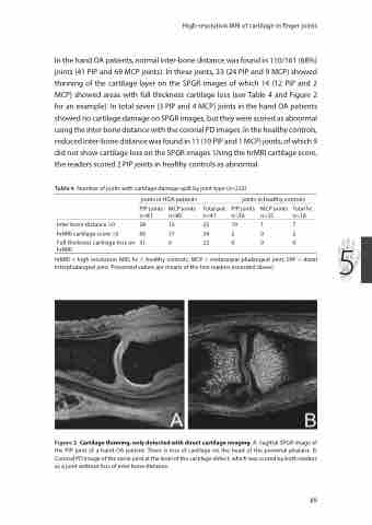
Figure 2. Cartilage thinning, only detected with direct cartilage imaging. A: Sagittal SPGR image of the PIP joint of a hand OA patient. There is loss of cartilage on the head of the proximal phalanx. B: Coronal PD image of the same joint at the level of the cartilage defect, which was scored by both readers as a joint without loss of inter bone distance.
89
