
Page 109 - Imaging of Osteoarthritis and Rheumatoid Arthritis in Hand Joints
P. 109
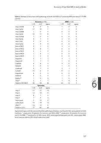
 Table 2. Number of structures with pathology on both low field 0.2T extremity MRI and clinical 1.5T MRI scanner.
Erosions (n=40)
1.5T 0.2T agree
mcp 2 distal 000 mcp 2 prox 17107 mcp 3 distal 100 mcp 3 prox 12118 mcp 4 distal 100 mcp 4 prox 430 mcp 5 dist 000 mcp 5 prox 944 base of MC1 432 base of MC2 481 base of MC3 030 base of MC4 141 base of MC5 120 trapezius 211 trapezoid 111 Capitate 885 Hamate 000 scaphoid 975 Lunate* 1165 triquetrum 865 Pisiform 000 Radius** 100 Ulna** 211 Total (n=907/867) 96 78 46
BME (n=39)
1.5T 0.2T agree 100
120
000
100
000
200
000
000
321
000
000
000
000
311
100
400
100
511
11 2 2
600
100
100 6 100
42 8 5
mcp 2
mcp 3
mcp 4
mcp 5 intercarpal radiocarpal DRU***
Total (n=273)
Synovitis
1.5T 0.2T agree 17 17 15 12 14 11 8 7 6
7 4 3
5 10 4 16 19 13 12 12 8 77 83 60
Agreement represents the amount that the pathological features was found in the same patient on both machines. * analysed in 39 patients for erosions and 38 for BME ** analysed in 34 patients for erosions and 33 for BME, ***analysed in 33 DRU-joints. MCP, metacarpal phalangeal joint; MC, metacarpal; BME, bone marrow edema; DRU, distal radio-ulnar joint
Accuracy of low field MRI in early arthritis
107
