
Page 54 - Magnesium-based supports for stem cell therapy of vascular disease - Mónica Echeverry Rendón
P. 54
CHAPTER 3
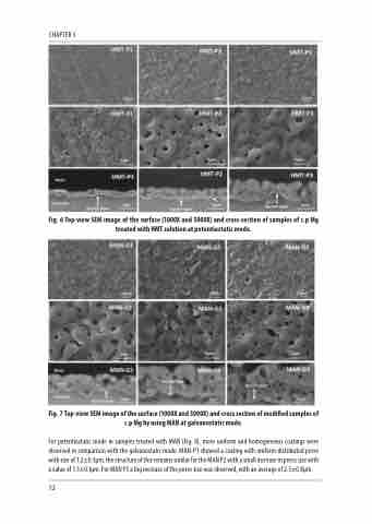
Fig. 6 Top-view SEM image of the surface (1000X and 5000X) and cross-section of samples of c.p Mg treated with HMT solution at potentiostatic mode.
Fig. 7 Top-view SEM image of the surface (1000X and 5000X) and cross section of modified samples of c.p Mg by using MAN at galvanostatic mode.
For potentiostatic mode in samples treated with MAN (Fig. 8), more uniform and homogeneous coatings were observed in comparison with the galvanostatic mode. MAN-P1 showed a coating with uniform distributed pores with size of 1.2±0.3μm, the structure of this remains similar for the MAN P2 with a small increase in pores size with a value of 1.5±0.1μm. For MAN P3 a big increase of the pores size was observed, with an average of 2.5±0.8μm.
52
