
Page 50 - Human Bile Acid Metabolism: a Postprandial Perspective
P. 50
Chapter 3
in the same order during the experiment: A, P, H, V, R. At 09:00 h, t = 0, the pigs received their test meal via the gastric feeding tube. For a pig of 25 kg, the test meal consisted a 600 mL mixture of 78 g of crude whey protein isolate (100% Premium Whey Protein; Body Fortress, Bohemia, NY) and 110 g of carbohydrates (Malto dextrin; Muscle Feast, Hebron, OH) in water, and 22 g of olive oil (30% of daily energy intake; Korger, Cincinnati, OH). We administered the complete test meal within approximately 5 min and took postprandial blood samples at t = 0, 10, 20, 30, 45, 60, 90, 120, 180 and 240 min. After the experiment, pigs were returned to their normal cages with their normal food and water available. Malfunctioning catheters accounted for missing data (two hepatic vein catheters and one renal vein catheter).
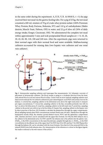
Fig. 1. Intravascular sampling catheters and transorgan flux measurements. (A) Schematic overview of placement of intravascular catheters. The blood stream is going in a clockwise direction from the heart with arterial blood in red and venous blood in blue. Circles with an arrow indicate sampling catheters and black arrows indicate the two infusion catheters for para-aminohippuric acid (PAH) and urinary loss via the kidneys. A: arterial line, sampling catheter in the abdominal aorta above the right renal artery to measure pre-organ concentrations; V: venous line, sampling catheter in the iliac circumflex profunda vein with its tip 5 cm above the bifurcation; R: renal line, sampling catheter in the left renal vein; P: portal line, sampling catheter in the portal vein with its tip in the liver hilus; H: hepatic line, sampling catheter in the hepatic vein by direct puncture of the liver. (B) Formulas to calculate the plasma flow through the organ compartments. Blood sampling started when PAH concentrations reached steady state (60 min). Plasma flow through the hindquarter muscle (HQ), the intestine (portal drained viscera, PDV) and the splanchnic compartment (SPL, intestine and liver) can be calculated with the upper flow formula. For HQ the PAH infusion site is the catheter in the abdominal aorta. For PDV and SPL the infusion site is the catheter into the splenic vein. [PAH]pre is the PAH concentration in the main bloodstream, sampled through line A. [PAH]post is the PAH concentration in the efferent vein of the organ: line P for PDV, line H for SPL and line V for HQ. Plasma flow through the kidneys (K) can be calculated with the lower formula. Note that for K the extracted PAH is the infused [PAH] from both infusion sites. (For interpretation of the references to colour in this figure legend, the reader is referred to the web version of this article.)
48
