
Page 66 - New epidemiological and PSMA-expression based paradigms in salivary gland tumors
P. 66
Chapter 4
cfDNA Assay V1 , Thermo Fisher Scientific, Waltham, MA, USA). With a limit of detection (LOD) down to 0.3% no indication was obtained for subclonal presence of these TP53 mutations in the three tumor samples.
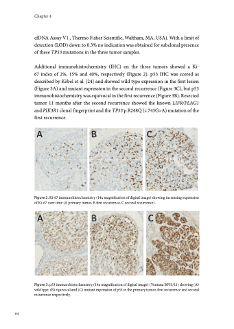
Additional immunohistochemistry (IHC) on the three tumors showed a Ki- 67 index of 2%, 15% and 40%, respectively (Figure 2). p53 IHC was scored as described by Köbel et al. [24] and showed wild type expression in the first lesion (Figure 3A) and mutant expression in the second recurrence (Figure 3C), but p53 immunohistochemistry was equivocal in the first recurrence (Figure 3B). Resected tumor 11 months after the second recurrence showed the known LIFR/PLAG1 and PIK3R1 clonal fingerprint and the TP53 p.R248Q (c.743G>A) mutation of the first recurrence.
Figure 2. Ki-67 immunohistochemistry (14x magnification of digital image) showing increasing expression of Ki-67 over time (A primary tumor, B first recurrence, C second recurrence).
Figure 3. p53 immunohistochemistry (14x magnification of digital image) (Ventana BP53/11) showing (A) wild type, (B) equivocal and (C) mutant expression of p53 in the primary tumor, first recurrence and second recurrence respectively.
64
