
Page 18 - New epidemiological and PSMA-expression based paradigms in salivary gland tumors
P. 18
16
Chapter 1
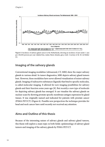
Figure 5: Incidence of salivary gland cancer in the Netherlands, showing an incidence of just under 1 case per 100,000 persons per year (dashed line, males; black, females; grey, total). Courtesy of dr. M. de Ridder [5].
Imaging of the salivary glands
Conventional imaging modalities (ultrasound, CT, MRI) show the major salivary glands in various detail. In tumor diagnostics, MRI depicts salivary gland tumors best. However, these modalities have never allowed visualization of minor salivary glands. Imaging of radioactive substances (ligands) that bind to specific molecules, is called molecular imaging. It allowed for new imaging possibilities for salivary glands and their function some years ago [8]. But recently a new type of molecule for depicting salivary glands has emerged. It can visualize the salivary glands on nuclear scans by showing prostate specific membrane antigen expression by gland tissues. It was originally named and indicated for patients with prostate cancer (PSMA PET/CT) (Figure 6). Possible new perspectives the technique provides for head and neck cancer have until recently not received any attention.
Aims and Outline of this thesis
Because of the interesting nature of salivary glands and salivary gland tumors, this thesis will explore a main topic in both fields: epidemiology of salivary gland tumors and imaging of the salivary glands by PSMA PET/CT.
