
Page 14 - New epidemiological and PSMA-expression based paradigms in salivary gland tumors
P. 14
12
Chapter 1
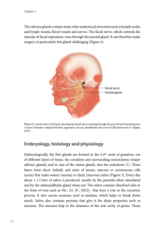
The salivary glands contain many other anatomical structures such as lymph nodes and lymph vessels, blood vessels and nerves. The facial nerve, which controls the muscles of facial expression, runs through the parotid gland. It can therefore make surgery of particularly this gland challenging (Figure 2).
Figure 2: Lateral view of the head, showing the facial nerve running through the parotid and branching into 5 major branches: temporal/frontal, zygomatic, buccal, mandibular and cervical (Illustration by R. Slagter, 2021).
Embryology, histology and physiology
Embryologically, the first glands are formed in the 4-6th week of gestation, out of different layers of tissue, the ectoderm and surrounding mesenchyme (major salivary glands) and in case of the minor glands, also the endoderm [1]. These layers form ducts (tubuli) and units of serous, mucous or seromucous cells (acini) that make watery (serous) or slimy (mucous) saliva (Figure 3). Every day about 1-1.5 liter of saliva is produced, mostly by the parotids when stimulated and by the submandibular gland when not. The saliva contains dissolved salts in the form of ions such as Na+, Cl-, K+, HCO3- that have a role in the excretion process. It also carries enzymes such as amylase, which helps to break down starch. Saliva also contains proteins that give it the slimy properties such as mucines. The mucines help in the clearance of the oral cavity of germs. These
Facial nerve Parotid gland
