
Page 134 - New epidemiological and PSMA-expression based paradigms in salivary gland tumors
P. 134
132
Chapter 8
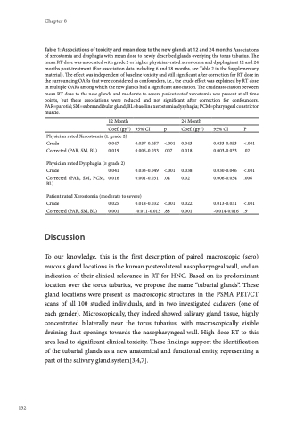
Table 1: Associations of toxicity and mean dose to the new glands at 12 and 24 months Associations of xerostomia and dysphagia with mean dose to newly described glands overlying the torus tubarius. The mean RT dose was associated with grade 2 or higher physician-rated xerostomia and dysphagia at 12 and 24 months post-treatment (For association data including 6 and 18 months, see Table 2 in the Supplementary material). The effect was independent of baseline toxicity and still significant after correction for RT dose in the surrounding OARs that were considered as confounders, i.e., the crude effect was explained by RT dose in multiple OARs among which the new glands had a significant association. The crude association between mean RT dose to the new glands and moderate to severe patient-rated xerostomia was present at all time points, but these associations were reduced and not significant after correction for confounders. PAR=parotid; SM=submandibular gland; BL=baseline xerostomia/dysphagia; PCM=pharyngeal constrictor muscle.
12 Month
Coef. (gy-1) Physician rated Xerostomia (≥ grade 2)
Crude 0.047 Corrected (PAR, SM, BL) 0.019
Physician rated Dysphagia (≥ grade 2)
Crude 0.041
Corrected (PAR, SM, PCM, 0.016 BL)
24 Month
Patient rated Xerostomia (moderate to severe)
Crude 0.025 0.018-0.032 Corrected (PAR, SM, BL) 0.001 -0.011-0.013
Discussion
95% CI
0.037-0.057 0.005-0.033
0.033-0.049 0.001-0.031
p Coef.
<.001 0.043 .007 0.018
<.001 0.038 .04 0.02
<.001 0.022 .88 0.001
(gy-1)
95% CI P
0.033-0.053 <.001 0.003-0.033 .02
0.030-0.046 <.001 0.006-0.034 .006
0.013-0.031 <.001 -0.014-0.016 .9
To our knowledge, this is the first description of paired macroscopic (sero) mucous gland locations in the human posterolateral nasopharyngeal wall, and an indication of their clinical relevance in RT for HNC. Based on its predominant location over the torus tubarius, we propose the name “tubarial glands”. These gland locations were present as macroscopic structures in the PSMA PET/CT scans of all 100 studied individuals, and in two investigated cadavers (one of each gender). Microscopically, they indeed showed salivary gland tissue, highly concentrated bilaterally near the torus tubarius, with macroscopically visible draining duct openings towards the nasopharyngeal wall. High-dose RT to this area lead to significant clinical toxicity. These findings support the identification of the tubarial glands as a new anatomical and functional entity, representing a part of the salivary gland system[3,4,7].
