
Page 131 - New epidemiological and PSMA-expression based paradigms in salivary gland tumors
P. 131
Tubarial salivary glands: A potential new organ at risk for radiotherapy
cytoplasmic expression of PSMA with a luminal preference, comparable to the mucous aspect and PSMA-ligand uptake of minor salivary glands in the palate (Fig.3; Fig.1 in the Supplementary material). There was no amylase expression in the gland cells consistent with very low numbers of serous acini, similar to the sublingual glands. The 3D histology reconstruction illustrates the anatomical distribution of glandular tissue and draining ducts (Fig.4; interactive 3D-PDF Fig.2 in the Supplementary material). MRI images of a healthy volunteer showed a subtle tissue structure with lower signal intensity on the T2 sequence, compatible with glandular tissue, was identified at the expected location of the tubarial gland on the medial side of the torus tubarius (Fig.3 in the Supplementary material). Small T2-intense dots were present within this tissue structure, which may represent the macroscopic duct openings seen in the cadavers and 3D histology reconstruction.
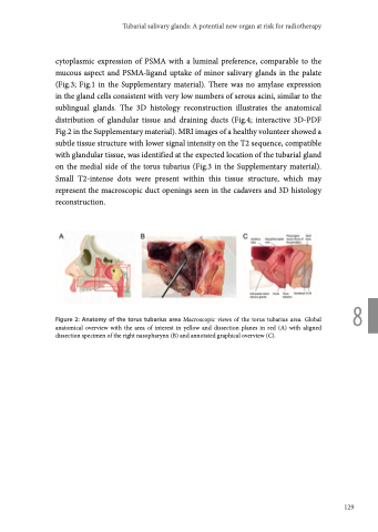
Figure 2: Anatomy of the torus tubarius area Macroscopic views of the torus tubarius area. Global 8 anatomical overview with the area of interest in yellow and dissection planes in red (A) with aligned
dissection specimen of the right nasopharynx (B) and annotated graphical overview (C).
129
