
Page 127 - New epidemiological and PSMA-expression based paradigms in salivary gland tumors
P. 127
Tubarial salivary glands: A potential new organ at risk for radiotherapy
Introduction
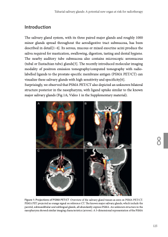
The salivary gland system, with its three paired major glands and roughly 1000 minor glands spread throughout the aerodigestive tract submucosa, has been described in detail[1–4]. Its serous, mucous or mixed exocrine acini produce the saliva required for mastication, swallowing, digestion, tasting and dental hygiene. The nearby auditory tube submucosa also contains microscopic seromucous (tubal or Eustachian tube) glands[5]. The recently introduced molecular imaging modality of positron emission tomography/computed tomography with radio- labelled ligands to the prostate-specific membrane antigen (PSMA PET/CT) can visualize these salivary glands with high sensitivity and specificity[6]. Surprisingly, we observed that PSMA PET/CT also depicted an unknown bilateral structure posterior in the nasopharynx, with ligand uptake similar to the known major salivary glands (Fig.1A; Video 1 in the Supplementary material).
8
Figure 1: Projections of PSMA PET/CT Overview of the salivary gland tissues as seen on PSMA PET/CT. PSMA PET projected as orange signal on reference CT. The known major salivary glands, which include the parotid, submandibular and sublingual glands, all abundantly express PSMA. An unknown structure in the nasopharynx showed similar imaging characteristics (arrows). A 3-dimensional representation of the PSMA
125
