
Page 42 - Synthesis of Functional Nanoparticles Using an Atmospheric Pressure Microplasma Process - LiangLiang Lin
P. 42
Chapter 2
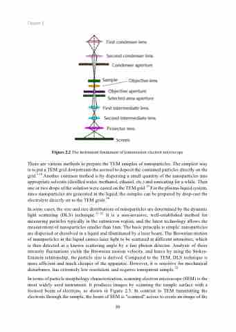
Figure 2.2 The instrument fundament of transmission electron microscope
There are various methods to prepare the TEM samples of nanoparticles. The simplest way is to put a TEM grid downstream the aerosol to deposit the contained particles directly on the grid.1,18 Another common method is by dispersing a small quantity of the nanoparticles into appropriate solvents (distilled water, methanol, ethanol, etc.) and sonicating for a while. Then one or two drops of the solution were casted on the TEM grid.19 For the plasma-liquid system, since nanoparticles are generated in the liquid, the samples can be prepared by drop-cast the electrolyte directly on to the TEM grids.20
In some cases, the size and size distributions of nanoparticles are determined by the dynamic light scattering (DLS) technique.21–23 It is a non-invasive, well-established method for measuring particles typically in the submicron region, and the latest technology allows the measurement of nanoparticles smaller than 1nm. The basic principle is simple: nanoparticles are dispersed or dissolved in a liquid and illuminated by a laser beam. The Brownian motion of nanoparticles in the liquid causes laser light to be scattered at different intensities, which is then detected at a known scattering angle by a fast photon detector. Analysis of these intensity fluctuations yields the Brownian motion velocity, and hence by using the Stokes- Einstein relationship, the particle size is derived. Compared to the TEM, DLS technique is more efficient and much cheaper of the apparatus. However, it is sensitive for mechanical disturbance, has extremely low resolution, and requires transparent sample.24
In terms of particle morphology characterization, scanning electron microscope (SEM) is the most widely used instrument. It produces images by scanning the sample surface with a focused beam of electrons, as shown in Figure 2.3. In contrast to TEM transmitting the electrons through the sample, the beam of SEM is "scanned" across to create an image of the
30
